Yes, vision therapy can meaningfully support recovery after a stroke or neurological condition. Many survivors experience visual disruptions — double vision, tracking problems, or visual field loss — that standard eye exams often miss. Neuro-optometric rehabilitation, a specialized branch of vision therapy, addresses these issues directly by retraining the brain-eye connection through targeted exercises and clinical interventions.
A stroke changes more than movement and speech. For a significant portion of survivors, the visual system is quietly disrupted in ways that slow recovery, interfere with reading, and make daily tasks unexpectedly difficult. The frustrating part is that many of these visual problems go unidentified during acute care, leaving patients struggling without a clear explanation for what they are experiencing.
Key Takeaways
- Stroke and traumatic brain injury (TBI) frequently damage the visual pathways, causing problems like double vision, convergence insufficiency, and hemianopia (partial visual field loss).
- Neuro-optometric rehabilitation uses targeted exercises and prism lenses to retrain the brain-eye system — it is not the same as standard vision therapy for children with reading difficulties.
- Many visual symptoms after a stroke are neurological in origin, meaning a routine eye exam will not detect them. A functional eye exam is required for accurate assessment.
- Early intervention appears to produce better outcomes, but recovery of visual function has been documented months and even years post-stroke.
- Common overlooked symptoms include light sensitivity, visual overwhelm symptoms, and difficulty with balance or spatial awareness.
- Not every provider offers neuro-optometric care — seeking a behavioural or neuro-optometrist is an important distinction when looking for the right specialist.
Why Does a Stroke Affect Vision in the First Place?
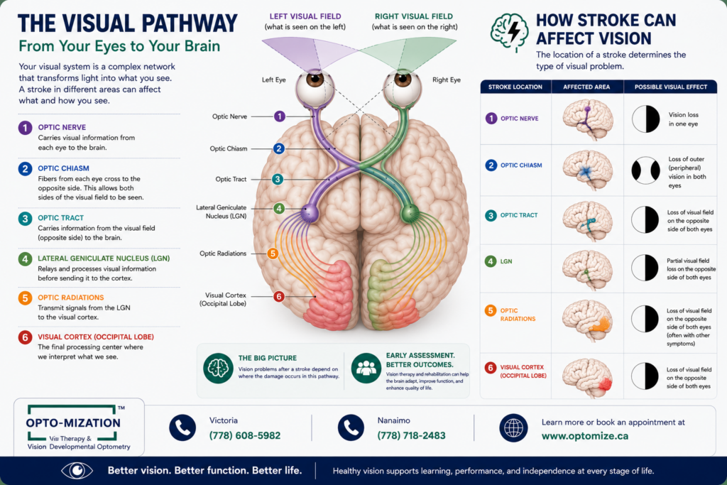
The visual system is not confined to the eyes. A large portion of the brain is dedicated to processing what we see, and nearly every stroke location has the potential to disrupt some part of that process. The occipital lobe handles visual processing, the parietal lobe manages spatial awareness, and the cerebellum coordinates eye movements. Damage to any of these areas produces distinct visual symptoms.
Research suggests that up to 65% of stroke survivors experience some form of visual impairment, yet vision is often the last system assessed during hospital rehabilitation. This creates a gap where patients are discharged with unaddressed visual dysfunction that directly limits their ability to participate in physical and cognitive therapy.
What makes this especially tricky is that visual symptoms after neurological events don’t always look like “eye problems.” A person may report feeling dizzy in grocery stores, struggling to read despite having new glasses, or feeling like they are bumping into things on one side. These are neurological vision symptoms, not optical ones.
What Visual Problems Commonly Occur After a Stroke?
| Visual Problem | What It Feels Like | Underlying Cause |
| Hemianopia | Missing half the visual field on one side | Damage to the visual cortex or optic radiations |
| Diplopia (double vision) | Seeing two overlapping images | Disrupted coordination of eye muscles from brain stem damage |
| Convergence insufficiency | Difficulty focusing on near objects; words blur when reading | Impaired binocular coordination |
| Visual neglect | Ignoring stimuli on the affected side, even with full visual field | Parietal lobe damage affecting attention |
| Photophobia | Overwhelming sensitivity to light indoors and outdoors | Disrupted neural filtering of light input |
| Oculomotor dysfunction | Difficulty tracking moving objects or shifting gaze accurately | Cerebellar or frontal lobe involvement |
Some of these conditions, like convergence insufficiency, respond very well to structured therapy. Others, like established hemianopia, may be partially compensated through specific scanning strategies and prism adaptation. The key word is “addressed” rather than “cured” — the goal of neuro-optometric rehabilitation is functional improvement, not always anatomical reversal.
How Is Neuro-Optometric Vision Therapy Different from Regular Vision Therapy?
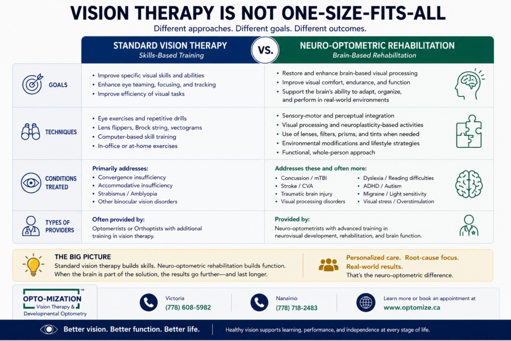
Standard vision therapy — the kind used to treat children with convergence problems or amblyopia — focuses on training the visual system when the brain itself is structurally intact. Neuro-optometric rehabilitation is designed for cases where the brain has been directly injured. The approach, the exercises, and the clinical reasoning are substantially different.
Neuro-optometrists complete additional post-graduate training specifically in acquired brain injury and neurological vision disorders. The College of Optometrists in Vision Development (COVD) and the Neuro-Optometric Rehabilitation Association (NORA) both offer fellowship designations that indicate advanced competency in this area.
In practice, this means the therapist is thinking about neuroplasticity rather than muscle strengthening alone. The brain’s capacity to form new pathways doesn’t stop after an injury, and this is precisely what rehabilitation exploits. Sessions might combine vergence exercises with balance training, or use yoked prisms to shift the patient’s entire spatial map and reduce visual midline shift syndrome.
Step-by-Step: What to Expect From Neuro-Optometric Rehabilitation
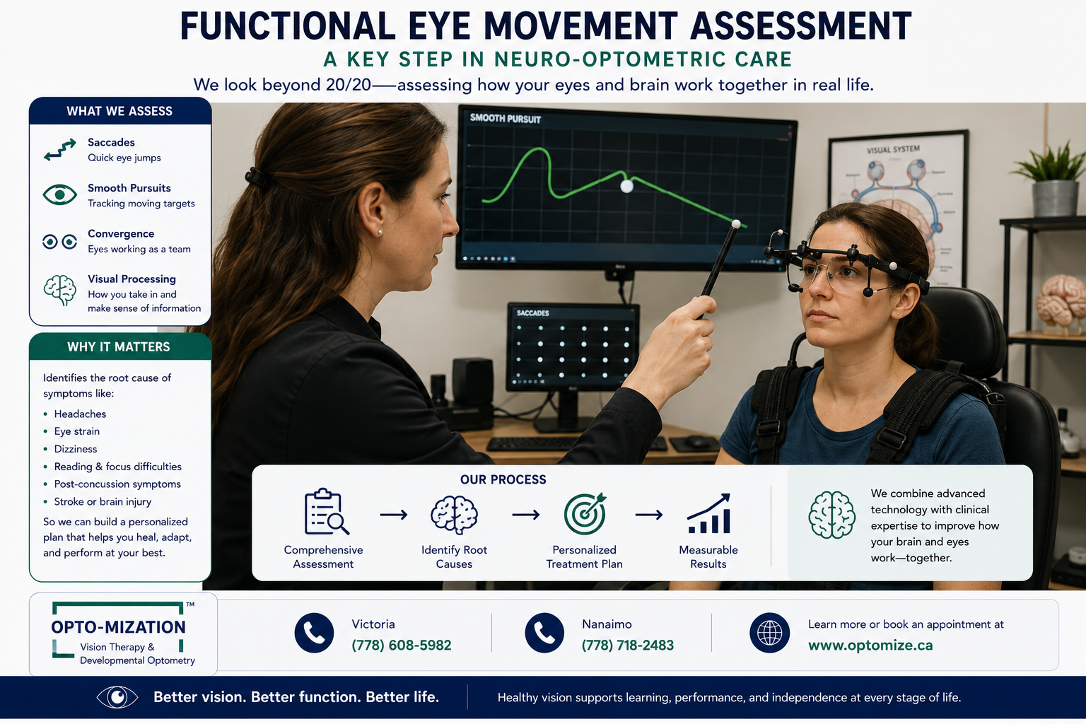
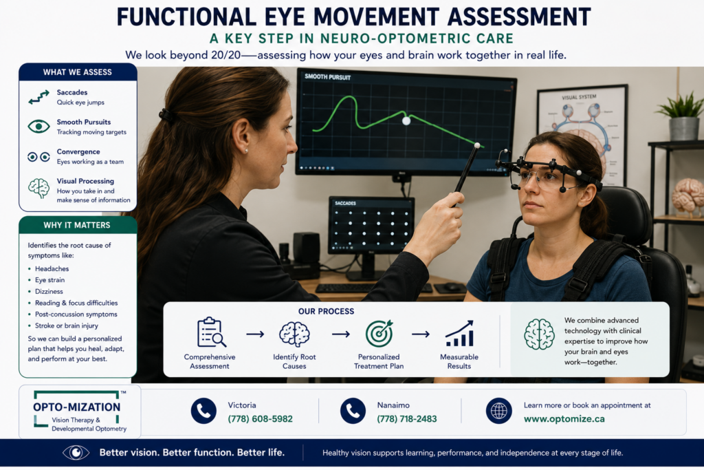
- Comprehensive functional vision assessment: This is far more detailed than a standard eye exam. The clinician evaluates eye teaming, tracking accuracy, visual field integrity, visual processing speed, and how your vision interacts with balance and posture. If you have never had a functional eye exam, this is where the process begins.
- Identify which deficits are primary vs. secondary: Some symptoms (like reading difficulty) are caused by a primary deficit (convergence insufficiency), while others are compensatory patterns the brain developed in response to the initial injury. Treating the right root cause matters.
- Develop an individualized therapy plan: No two stroke survivors present identically. A plan might include in-office weekly sessions, home exercises, therapeutic lenses or prisms, and coordination with other therapists (occupational, physical, or speech).
- Progress through graded challenges: Exercises increase in complexity as the visual system adapts. Early sessions may focus on stable, single-plane targets. Later sessions introduce movement, divided attention, and real-world simulations like reading text in a noisy environment.
- Re-evaluate and adjust: Most programs include formal progress assessments at regular intervals. Objective measurements — not just self-report — guide decisions about continuing, modifying, or concluding therapy.
What Does the Evidence Actually Say?
The evidence base for neuro-optometric rehabilitation is growing but still maturing. Randomized controlled trials on post-stroke vision therapy are limited, partly because the patient population is heterogeneous and outcomes are difficult to standardize. However, studies on specific conditions; particularly convergence insufficiency, diplopia management with prisms, and compensatory scanning training for hemianopia, show consistent evidence of functional improvement.
For traumatic brain injury (TBI), which shares many visual symptoms with stroke, the research is more extensive. A landmark study by Ciuffreda and colleagues found that approximately 90% of TBI patients have some measurable visual dysfunction, and structured vision rehabilitation produced significant improvement in reading, balance, and quality of life measures for a majority of participants.
The honest clinical picture is this: vision therapy after a stroke is not a guaranteed cure, and results vary based on the location and severity of injury, the patient’s age and general health, how much time has passed since the stroke, and the quality and consistency of the rehabilitation program. But “results vary” is very different from “it doesn’t work.” Many patients experience meaningful, life-altering improvements.
Symptoms That Shouldn’t Be Ignored After a Stroke
One of the most important things rehabilitation specialists observe is how often visual symptoms get attributed to fatigue or aging rather than neurological injury. A few patterns that consistently warrant evaluation:
- Persistent light sensitivity that wasn’t present before the event
- Difficulty reading despite a current glasses prescription — especially if the problem is losing your place on a line rather than blurry letters
- Feeling off-balance or dizzy in visually busy environments, which can be a sign of visual overwhelm symptoms
- A sense of pressure behind eyes or persistent eye strain without a clear cause
- Bumping into objects on one side or missing food on one side of the plate
- Discomfort or double vision when watching television or using a screen
If any of these symptoms appeared after a stroke, TBI, or other neurological event, they are not likely to resolve on their own, and they may be addressable with the right specialist assessment.
Common Mistakes in Post-Stroke Vision Care
- Relying solely on a standard optometry exam: A routine refraction checks clarity and eye health, but it does not evaluate binocular function, tracking, visual processing, or field deficits linked to brain injury. Without the right test, the right diagnosis cannot happen.
- Waiting too long before seeking help: Neuroplasticity is most active in the early months post-stroke, but it doesn’t shut off. Still, earlier intervention tends to yield faster results and prevents compensatory habits (like head-turning to compensate for a visual field loss) from becoming deeply ingrained.
- Treating vision in isolation: Visual rehabilitation works best as part of a coordinated care approach. A neuro-optometrist who communicates with an occupational therapist or physiatrist will produce better functional outcomes than one working in isolation.
- Assuming nothing can be done: Patients are sometimes told their visual problems are permanent. In some cases this is accurate. In many others, it reflects a lack of familiarity with neuro-optometric options, not the actual limits of recovery.
- Stopping therapy too early: Visual rehabilitation often follows a non-linear trajectory. Patients may plateau temporarily before making new gains. Consistent attendance and home exercise compliance significantly affect long-term outcomes.
What About Other Neurological Conditions?
Vision therapy isn’t limited to stroke recovery. Neuro-optometric rehabilitation is also used for individuals with acquired brain injury from car accidents or sports concussions, multiple sclerosis, Parkinson’s disease, post-COVID neurological symptoms, and developmental or acquired learning-related visual processing disorders.
| Condition | Common Visual Symptoms | Potential Neuro-Optometric Interventions |
| Concussion / mTBI | Blurred vision, headaches, sensitivity to light | Vergence therapy, tinted lenses, vestibular-visual integration |
| Multiple sclerosis | Optic neuritis, oscillopsia, diplopia | Prism lenses, gaze stabilization, fatigue management strategies |
| Parkinson’s disease | Reduced blink rate, convergence problems, contrast sensitivity loss | Prismatic correction, saccadic training, ambient lenses |
| Post-COVID syndrome | Brain fog affecting visual attention, eye strain, photophobia | Functional vision assessment, graded visual activity exposure |
The common thread across all these conditions is that the visual system is affected not at the eye level, but at the processing level. Standard optical corrections cannot address that. Knowing how often to get re-evaluated as your condition changes is also worth discussing with your provider. Questions like how often should you get an eye exam become more nuanced when you’re managing a neurological condition.
Frequently Asked Questions
Can the brain heal after a stroke?
Yes, though the extent of healing depends on the severity and location of the injury, the patient’s age, and the quality of rehabilitation. The brain undergoes neuroplasticity, a process of forming new neural connections to compensate for damaged areas. This process is most active in the first three to six months post-stroke but continues for years. Rehabilitation therapies, including vision therapy, are designed to actively leverage and accelerate this process.
What is vision therapy after a stroke?
Post-stroke vision therapy, more precisely called neuro-optometric rehabilitation, is a structured program of exercises and clinical interventions designed to retrain the brain-eye connection after neurological injury. It is conducted by a neuro-optometrist or behavioural optometrist with specialized training, and it addresses issues like double vision, tracking problems, visual field deficits, and balance disorders with a visual origin. Sessions typically occur weekly in-clinic and are supplemented with home exercises.
What is the best treatment for stroke?
Stroke treatment is multi-layered and depends heavily on the type and severity of the event. In acute care, clot-busting medications (tPA) or mechanical thrombectomy are used when eligible patients are treated quickly. In rehabilitation, the strongest evidence supports intensive, task-specific therapy across physical, occupational, speech, and cognitive domains, started as early as medically stable. Vision rehabilitation is increasingly recognized as a critical but often missing component of comprehensive stroke recovery.
What to avoid after a stroke?
In the recovery period, stroke survivors should avoid activities that increase the risk of a second stroke — this includes uncontrolled hypertension, smoking, excessive alcohol, and sedentary behavior. From a visual rehabilitation standpoint, patients should avoid dismissing visual symptoms as minor or age-related, driving before their visual function has been properly assessed, and skipping rehabilitation appointments, as consistency is directly linked to outcomes. Avoid making assumptions about what is and isn’t treatable without a specialist evaluation.
What fruit is best for stroke?
No single fruit prevents or treats stroke, but a diet rich in potassium, antioxidants, and fiber supports vascular health and reduces stroke risk factors. Citrus fruits like oranges and grapefruit are associated with reduced stroke risk in some observational studies, likely due to their flavonoid content. Berries (blueberries, strawberries) are high in anthocyanins linked to improved endothelial function. For post-stroke recovery, nutritional support should be discussed with a registered dietitian as part of a comprehensive care plan.
Conclusion: Visual Recovery Is Possible, But You Need the Right Support
Stroke survivors are often told to expect a plateau in their recovery, usually within the first year. The visual system challenges that assumption. With the right assessment and a structured neuro-optometric rehabilitation program, meaningful functional improvement is achievable well beyond that window.
The most important first step is getting evaluated by someone who actually looks at all the ways your visual system functions, not just your prescription. Standard eye care, as thorough as it may be, wasn’t designed to detect or treat neurological visual dysfunction. That gap is exactly what specialized providers exist to fill.
If you or someone you care for is experiencing visual difficulties following a stroke, brain injury, or another neurological condition, Opto-Mization offers neuro-optometric assessments designed specifically for these situations. Their team understands the difference between optical problems and neurological ones, and they work with patients and their care teams to build a meaningful rehabilitation path. Reach out directly at (778) 608-5982 to schedule a functional vision assessment and take a concrete step toward visual recovery.