
Floaters, flashes, and cobweb-like shapes in your vision are usually caused by age-related changes inside the vitreous, the gel-like fluid that fills your eye. Most are harmless and fade from your conscious awareness over time. However, a sudden increase in floaters, flashes of light, or a shadow across your vision can signal a retinal tear or detachment, which requires prompt professional evaluation.
Introduction
You glance up at a bright sky or a plain white wall and notice something drifting across your field of view, a wispy thread, a transparent blob, or something that looks almost like a cobweb floating just out of reach. You move your eyes to look directly at it, and it moves too. This is one of the more unsettling visual experiences people encounter, and it sends a lot of people searching for answers.
The good news is that the vast majority of floaters are benign. The less reassuring truth is that some presentations genuinely do warrant a same-day eye exam. Understanding the difference matters.
Key Takeaways
- Floaters are shadows cast by clumps or strands inside the vitreous gel of your eye, not particles on the surface of your eye or lens.
- Most floaters are a normal, age-related phenomenon called posterior vitreous detachment (PVD) and are not dangerous on their own.
- Flashes of light occurring alongside floaters can indicate traction on the retina, which increases the risk of a retinal tear.
- A sudden shower of new floaters, particularly with flashes or a dark curtain effect, is an eye emergency that requires same-day assessment.
- Routine eye exams are the only reliable way to determine whether floaters are harmless or linked to a structural problem at the back of the eye.
- Most people adapt to benign floaters over time, but treatment options do exist for cases where floaters significantly affect quality of vision.
Things You Must Know
1. “New” is the word that changes everything.
If you have noticed a handful of floaters for years and they have stayed roughly the same, that is a very different situation from waking up one morning to find a sudden storm of new dots, threads, or shapes you have never seen before. The suddenness of onset matters more than the number of floaters present. Any abrupt change in your floater pattern should be evaluated promptly, not monitored at home.
2. Floaters are not on your eye, they are inside it.
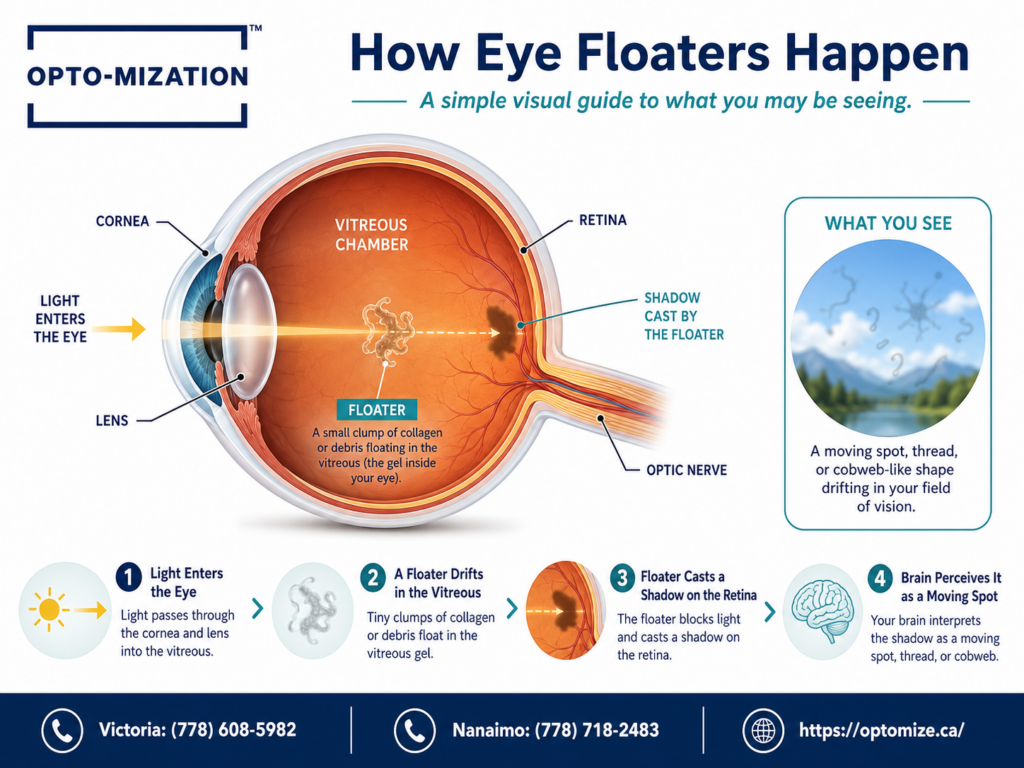
A common misconception is that floaters are something on the surface of the eye that can be rinsed away or blinked out. They are actually shadows projected onto your retina by debris suspended in the vitreous humor, the clear gel that fills the back chamber of your eye. This is why they drift lazily when your eyes move and why they seem to follow your gaze. No eye drops, supplements, or exercises have been shown to dissolve them reliably.
3. Floaters and light sensitivity can appear together, and both deserve attention.
Some people experiencing floaters also notice increased sensitivity to bright lights or visual disturbances in bright environments. If you are also dealing with light sensitivity that feels unusually intense, it is worth mentioning both symptoms to your optometrist, as the combination can sometimes point to inflammatory or pressure-related causes beyond simple vitreous changes.
What Exactly Are Eye Floaters?
The vitreous humor starts out as a smooth, transparent gel. As you age, the vitreous gradually liquefies and begins to shrink. Tiny fibers within the gel can clump together, and these clumps cast shadows on the retina as light passes through. What you perceive as a floater is actually that shadow, not the clump itself.
The shapes people describe vary widely: dots, specks, squiggly lines, threads, rings, and the classic cobweb or net-like structure. The cobweb appearance is particularly common during or after a posterior vitreous detachment, when the vitreous peels away from the retina and bunches of gel fibers gather into a loose, lacy structure near the center of your visual field.
Why Do Floaters Look Like Cobwebs Specifically?
The cobweb pattern is one of the most commonly reported floater types, and it tends to alarm people more than simple dots. It usually means the vitreous gel has partially detached from the back of the eye and collapsed into itself, with gel strands folding into a loosely connected mesh structure.
This is often the hallmark of a posterior vitreous detachment, which is considered a normal aging event. PVD typically occurs between the ages of 50 and 75, though it can happen earlier in people who are highly myopic (nearsighted), have had eye trauma, or have undergone certain eye surgeries.
Posterior vitreous detachment is estimated to affect the majority of people over the age of 70, with a significant percentage experiencing it in at least one eye by their mid-50s. For most of them, the event is harmless, though it can be accompanied by a brief increase in floaters and flashes.
What Causes Flashes of Light in Your Vision?
Flashes are different from floaters and carry a slightly more urgent connotation. They appear as brief streaks, arcs, or sparks of light, often described as looking like camera flashes or lightning in peripheral vision. They tend to appear in dim lighting or when the eyes move quickly.
The cause is mechanical. When the vitreous tugs on the retina, either during a PVD or due to traction in another context, it stimulates the retinal cells. Because those cells are designed to respond to light, the brain interprets the stimulation as a flash of light even though no actual light source caused it.
Flashes that are infrequent and tied to a known PVD episode are usually not dangerous. Flashes that are frequent, persistent, or appear alongside a sudden new wave of floaters raise concern for a retinal tear, since the vitreous may have pulled hard enough during detachment to create a small break in the retinal tissue.
When Are Floaters and Flashes a Warning Sign?
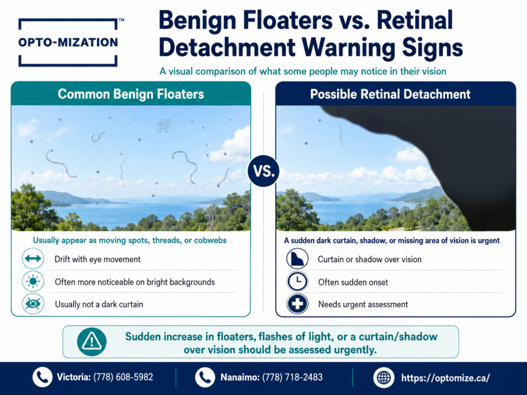
This is the question that matters most. Most floaters are benign, but certain presentations are considered red flags that warrant urgent evaluation, not a wait-and-see approach.
| Symptom | Likely Cause | Urgency Level |
| Occasional single floater, no change over time | Benign vitreous debris | Routine exam |
| Cobweb or ring-shaped floater, recent onset | Posterior vitreous detachment (PVD) | Prompt exam within days |
| Sudden shower of many new floaters | Possible retinal tear or bleed | Same-day evaluation |
| Flashes plus new floaters | Vitreous traction on retina | Same-day evaluation |
| Dark shadow or curtain in any part of vision | Retinal detachment | Emergency, go now |
What Happens During a Retinal Tear or Detachment?
If the vitreous pulls strongly enough as it detaches, it can tear the retina rather than simply separating cleanly from it. A retinal tear by itself is treatable, usually with laser photocoagulation or cryotherapy to seal the break and prevent it from progressing. Left undetected, a tear can allow fluid to seep beneath the retina, causing it to lift away from the underlying tissue.
Retinal detachment is a sight-threatening emergency. Once the retina separates, visual cells begin to lose their blood supply. The longer the detachment goes untreated, the greater the risk of permanent vision loss, particularly if the central area of the retina (the macula) becomes involved.
Retinal detachment affects approximately 1 in 10,000 people per year in the general population, but that rate rises meaningfully among those who have already experienced a posterior vitreous detachment, are highly myopic, or have a family history of retinal detachment.
Other Causes of Floaters and Visual Disturbances Worth Knowing
Not every floater comes from PVD. A few other conditions can produce similar symptoms:
- Diabetic retinopathy: Bleeding inside the eye from damaged retinal vessels can release blood cells into the vitreous, causing a sudden increase in dark floaters or a reddish haze.
- Uveitis: Inflammation inside the eye can produce floaters alongside redness, pain, and pressure behind the eyes, and it requires anti-inflammatory treatment rather than simple monitoring.
- Ocular migraines: These typically produce geometric, shimmering patterns (called an aura) that move across vision over 20 to 30 minutes. They are usually harmless and resolve on their own, but they are distinct from floaters caused by vitreous changes.
- Posterior capsule opacification: People who have had cataract surgery can develop clouding behind their artificial lens, which can produce visual disturbances that feel similar to floaters.
What to Expect at an Eye Exam for Floaters
If you visit an optometrist about floaters or flashes, here is the general process you can expect:
- Case history: Your optometrist will ask about when the floaters started, whether they changed suddenly, any flashes, and your general health and eye history.
- Visual acuity test: A standard check of how clearly you can see at distance and near.
- Dilated fundus examination: Eye drops are used to dilate your pupils, allowing a thorough view of the vitreous, retina, and optic nerve. This step is essential for ruling out retinal tears or detachment.
- Slit-lamp biomicroscopy: A high-magnification view of the structures inside the eye, including the vitreous gel.
- Additional imaging if needed: In some cases, optical coherence tomography (OCT) or ultrasonography may be used to get a more detailed picture of the retina and vitreous.
Because dilation is typically required, plan to have someone drive you or bring sunglasses, as your near vision and light sensitivity will be temporarily affected afterward.

Can Floaters Be Treated or Removed?
| Treatment Option | How It Works | Who It May Be Suitable For |
| Watchful waiting | Most people adapt as the brain learns to suppress floater shadows | Benign floaters with no retinal risk |
| YAG laser vitreolysis | A laser targets and vaporizes vitreous clumps | Large, vision-affecting floaters in suitable positions |
| Vitrectomy | Surgical removal of the vitreous gel and replacement with saline | Severe, debilitating floater cases; carries surgical risks |
YAG laser vitreolysis is not appropriate for all floater types. Its effectiveness and safety profile vary depending on the size and location of the vitreous opacity relative to the retina and lens, and patient selection is a critical factor in outcomes.
The reality for most patients is that treatment is not necessary. The brain is remarkably good at filtering out stable floaters over weeks to months. People often notice them far less intensely six months after their onset compared to how distracting they felt in the beginning.
Common Mistakes People Make When Dealing With Floaters
- Waiting too long to get checked after a sudden change. New floaters that appear abruptly need to be assessed quickly, not in a few weeks when a routine appointment becomes available.
- Assuming all floaters are the same. A single, long-standing floater and a sudden burst of dozens of new ones are entirely different situations clinically.
- Trying supplements or eye drops marketed for floaters. There is no well-established, clinically proven supplement regimen for eliminating vitreous floaters. Be cautious about products making these claims.
- Ignoring floaters alongside other symptoms. Floaters that come with eye pain, redness, or headaches deserve attention sooner rather than later, as that combination can indicate inflammation or elevated eye pressure.
- Skipping the dilated exam. A non-dilated exam cannot adequately assess the peripheral retina, which is where many tears originate.
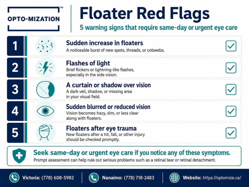
Studies examining patient behaviour after floater onset suggest a meaningful proportion of individuals delay seeking care by weeks or months, even when experiencing symptoms that could indicate a retinal tear. Early examination significantly improves outcomes when a tear is present.
Frequently Asked Questions
Why am I seeing cobwebs in my vision?
A cobweb-like shape drifting across your vision is almost always the result of a posterior vitreous detachment. As the vitreous gel peels away from the retina, strands of gel fiber collapse and fold into a loose, lacy structure. This structure floats in the vitreous chamber and casts a shadow on your retina that your brain registers as a cobweb or net-like shape. It typically becomes more noticeable against bright, evenly lit backgrounds like a pale sky or a white wall. A dilated eye exam can confirm whether the detachment is clean or associated with a retinal tear.
Are cobweb floaters serious?
For most people, cobweb floaters tied to a posterior vitreous detachment are not serious in themselves. The concern is whether the detachment occurred cleanly or caused a tear in the retinal tissue in the process. This distinction cannot be made without a professional dilated examination. A cobweb floater that appears suddenly, especially alongside flashes or a change in peripheral vision, should be evaluated promptly. If the exam confirms a clean PVD with no retinal damage, the floaters are typically harmless, though they may persist for some time.
Is it normal to see flashes with floaters?
Flashes accompanying floaters are common during an active posterior vitreous detachment, particularly in the weeks immediately after it starts. The flashes occur because the separating vitreous tugs on the retina, mechanically stimulating the photoreceptors and causing the brain to perceive light that is not actually there. This usually settles down as the vitreous fully detaches and stops pulling. Flashes that continue or worsen, especially with new floaters, suggest ongoing traction and should be reassessed. People who notice that their eyes struggle to adjust between distances alongside flashes should mention both issues at their exam.
How do you get rid of cobweb eye floaters?
For most people, cobweb floaters do not require active treatment. The brain adapts to stable floaters through a process called neurological suppression, where it gradually learns to filter them out of conscious awareness. For floaters that are genuinely disruptive to daily activities or central vision, a specialist may discuss YAG laser vitreolysis or, in more severe cases, vitrectomy surgery. Neither option is appropriate for everyone, and both carry risks. The first step is always a thorough eye exam to assess the floaters, confirm there is no retinal damage, and discuss realistic expectations for how the floaters may change over time.
Will floaters go away naturally?
Floaters rarely disappear entirely, but most people find them significantly less bothersome over time. Two things contribute to this: the floaters themselves may shift lower in the vitreous cavity, moving out of the central line of sight, and the brain becomes better at ignoring them through neurological adaptation. People with floaters stemming from a clear, uncomplicated PVD typically report much less interference with their vision after three to six months compared to initial onset. Floaters caused by bleeding, inflammation, or ongoing retinal issues behave differently and require management of the underlying condition.
Seeing Something Unusual in Your Vision? Get It Checked.
Floaters and flashes sit in an unusual middle ground: usually harmless, occasionally urgent, and impossible to assess accurately without a proper dilated eye exam. If your floaters are stable and have been present for a long time, a routine annual exam is the right approach. If something has changed recently, or if you are noticing flashes, a sudden increase in floaters, or any shadow in your visual field, that is not a situation to wait on.
The team at Opto-mization can perform a thorough dilated retinal examination to assess the health of your vitreous and retina and give you a clear picture of what is happening inside your eye. Call us at (778) 608-5982 to book your appointment. Catching a retinal tear early, when it is still a small, treatable break, is a very different situation from dealing with it after it has progressed.