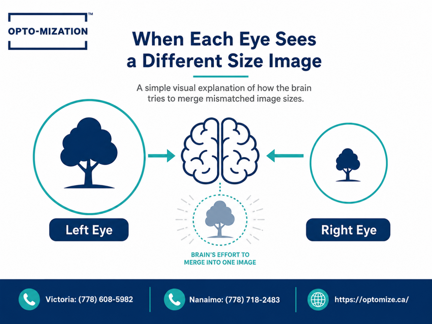
When objects appear larger through one eye than the other, the condition is called aniseikonia. It occurs when the two eyes send images of different sizes to the brain, disrupting binocular vision. This can result from differences in prescription, eye anatomy, or how the brain processes visual information, and it often warrants a functional vision assessment rather than a standard eye exam.
Introduction
Most people have tested it at some point: covering one eye, then the other, and noticing the world looks subtly different through each. For some, that difference is striking. Objects shift in perceived size, depth feels off, or reading becomes strangely uncomfortable without any obvious explanation.
This isn’t just a quirky optical illusion. When one eye consistently sees things larger or smaller than the other, it’s a measurable phenomenon with real effects on daily function, and it’s one that traditional eye exams often don’t catch.
Key Takeaways
- The condition where one eye perceives images as larger or smaller than the other is called aniseikonia.
- It can be caused by differences in prescription between eyes, lens design, eye anatomy, or neurological visual processing.
- Symptoms often include headaches, eyestrain, depth perception problems, and difficulty reading, even with a correct prescription.
- Standard eye exams typically don’t measure for aniseikonia; a functional vision assessment is usually needed.
- Treatment options range from specialized lens prescriptions to vision therapy, depending on the underlying cause.
- Left untreated, aniseikonia can interfere with school performance, work productivity, and quality of life.
Things You Must Know
1. A “Normal” Prescription Doesn’t Rule This Out
Many patients with aniseikonia have been told their eyesight is fine. That’s because standard visual acuity tests measure sharpness, not how the brain reconciles two slightly different images. You can see 20/20 in each eye individually and still experience significant discomfort because the brain is working overtime to merge mismatched images. If your eyes feel tired even when your prescription is correct, image size disparity between the eyes may be worth investigating.
2. The Difference Doesn’t Have to Be Large to Matter
Research suggests that an image size difference of even 1% between the two eyes can begin to cause symptoms in sensitive individuals. A 5% difference is generally considered clinically significant, but people experience discomfort across a wide range. This means the problem can be subtle on testing and still be the reason behind chronic headaches, poor depth perception, or difficulty concentrating on a page.
3. The Cause Affects the Treatment Path
Aniseikonia caused by a large prescription difference between eyes is treated differently than aniseikonia caused by neurological factors or post-surgical changes. Getting an accurate functional vision assessment before committing to a treatment approach matters, because the wrong lens design can actually make symptoms worse. A specialist who measures binocular vision function, not just refractive error, is better positioned to find the right solution.
What Is Aniseikonia and Why Does It Happen?
Aniseikonia (pronounced an-eye-seh-KOH-nee-uh) is the clinical term for a condition in which the images formed by each eye differ in size or shape. The brain receives two slightly different pictures and has to decide what to do with them.
Under normal circumstances, your visual system is remarkably good at blending the images from both eyes into a single, seamless view of the world. This process is called binocular fusion. When the images are too different in size, the brain struggles to fuse them, leading to suppression of one eye, eyestrain, distorted depth perception, or outright discomfort.
The Two Main Types
Optometrists generally distinguish between two forms. Optical aniseikonia arises from physical differences in the optical system itself, such as unequal prescriptions or lens choices. Neural aniseikonia comes from differences in how each eye’s signals are processed by the brain, which can occur after neurological events, concussions, or certain retinal conditions.
The distinction matters clinically because the two types respond to different interventions.
What Causes One Eye to See Things Larger or Smaller?
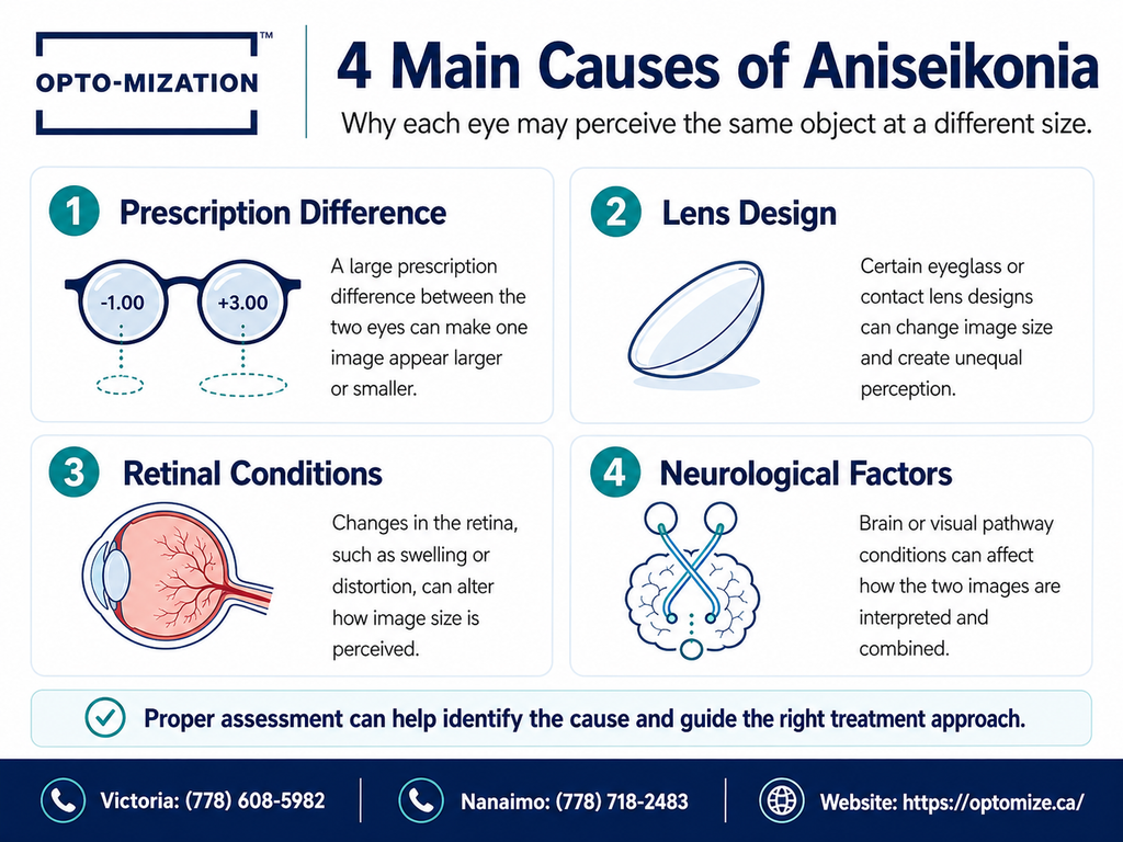
Several factors can create a meaningful size difference between the two eyes’ images.
Prescription Differences Between Eyes (Anisometropia)
When one eye needs a significantly stronger prescription than the other, the corrective lenses themselves can introduce image size differences. Spectacle lenses magnify or minify the image depending on the prescription, and when there is a large difference between the two lenses, the resulting image size disparity can be substantial enough to cause symptoms. This is one of the more common causes of induced aniseikonia.
Lens Type and Design
The choice of spectacle lens or contact lens affects magnification. Contact lenses, because they sit directly on the eye, generally produce less image size difference than spectacle lenses for the same prescription. High-powered or thick lenses can amplify size differences in ways that an off-the-shelf prescription doesn’t account for.
Retinal Conditions and Eye Surgery
Epiretinal membranes, macular conditions, and certain retinal surgeries can stretch or distort the photoreceptor arrangement in one eye, causing that eye to perceive images as a different size or shape even when the optical prescription is balanced. Post-surgical aniseikonia is well documented following procedures like cataract surgery with implanted lenses of different powers.
Neurological and Concussion-Related Changes
After a head injury or concussion, the brain’s ability to process and merge visual signals from both eyes can be disrupted. Some patients notice that objects look larger or smaller in one eye, or that the world looks tilted or unstable, starting only after a neurological event. This type of aniseikonia sits within the broader category of post-concussion vision syndrome and often requires a neuro-optometric approach to address effectively.
How the Brain Responds to Mismatched Images
The visual brain is adaptive, sometimes impressively so. When it receives two differently sized images, it has a few options: it can attempt to fuse them (resulting in eyestrain or headaches from the effort), suppress the input from one eye entirely, or allow both images to compete, which can cause instability or disorientation.
Long-term suppression of one eye’s input is particularly concerning in children, because it can lead to amblyopia (often called a lazy eye) if the brain chronically ignores one eye’s signal during the developmental years. For adults, suppression typically means reduced depth perception, difficulty judging distances, and fatigue during visually demanding tasks.
This is part of why people with undiagnosed aniseikonia sometimes struggle with tasks that require sustained visual attention. Reading, driving, and screen work all become more effortful when the brain is constantly working to reconcile two mismatched pictures. If you’re curious about related visual processing difficulties, the experience of busy patterns and crowded text overwhelming the visual system shares some underlying mechanisms with how the brain handles competing visual signals from two eyes.
Common Symptoms People Describe
- Headaches that worsen with reading or screen use
- Eyestrain even with a recent prescription
- Difficulty judging distances or depth
- Objects appearing tilted or distorted
- Double vision or ghosting, particularly when tired
- Dizziness or spatial disorientation
- Difficulty reading for extended periods
- Closing or covering one eye for relief
Many of these symptoms overlap with other binocular vision conditions, which is one reason an accurate assessment matters so much. The same cluster of complaints can have very different causes, and addressing the wrong one rarely helps.
People who find that their eyes feel tired even though their prescription is correct are often dealing with a binocular vision issue rather than a simple refractive one, and aniseikonia is one possibility worth ruling in or out.
How Is Aniseikonia Diagnosed?
Diagnosis requires specialized testing that goes beyond a standard refraction. A functional vision assessment can measure the actual perceived size difference between the two eyes using instruments and techniques designed specifically for this purpose.
- Case history and symptom review: A detailed discussion of when symptoms occur, how long they’ve been present, and what activities trigger them helps establish the clinical picture.
- Binocular vision testing: The clinician evaluates how the two eyes work together, including fusion ability, depth perception, and suppression patterns.
- Eikonometry: Specialized testing measures the degree of image size difference between the two eyes. This step is often skipped in routine eye exams.
- Retinal and neurological evaluation: If indicated, the assessment may include examination of the retina and consideration of neurological factors, particularly after head injury.
- Prescription analysis: The current prescription and lens design are reviewed to determine whether they may be contributing to or exacerbating the disparity.
What Are the Treatment Options?
| Treatment Approach | Best Suited For | How It Helps |
| Iseikonic (size) lenses | Optical aniseikonia from prescription differences | Specially designed lenses that equalize the perceived image size between the two eyes |
| Contact lenses | Patients with large prescription differences | Reduce magnification difference compared to spectacles |
| Vision therapy | Neural aniseikonia, post-concussion cases, suppression | Retrains binocular coordination and reduces suppression through structured exercises |
| Prism lenses | Associated alignment issues | Realign images to reduce fusion effort and symptom load |
| Treating the underlying condition | Retinal or neurological causes | Addressing the source condition may reduce or resolve the size disparity |
For patients whose aniseikonia developed after a concussion or neurological event, vision therapy is often a core part of the recovery process. It focuses on retraining how the brain processes and integrates the signals from both eyes, which can’t be corrected with lenses alone.
Studies have shown that vision therapy can significantly improve binocular coordination, reduce suppression, and alleviate symptoms in patients with binocular vision dysfunction, including cases where aniseikonia is part of the clinical picture. Outcomes tend to be better when treatment is started early and tailored to the specific pattern of dysfunction rather than applied generically.
How Does Aniseikonia Affect Daily Life?
| Activity | How Aniseikonia Can Interfere |
| Reading | Words may appear to shift, float, or be harder to track across a line |
| Screen work | Sustained near focus becomes fatiguing faster than expected |
| Driving | Depth perception issues can affect judging distances to other vehicles |
| Sports and athletics | Reduced spatial accuracy when catching, hitting, or tracking moving objects |
| Balance and movement | Visual instability can contribute to dizziness or unsteadiness |
| School performance (children) | Reading avoidance, difficulty copying from the board, apparent inattention |
Common Mistakes People Make With This Condition
- Assuming a new glasses prescription will fix it: If the underlying issue is how the brain processes the images, a prescription change alone often doesn’t resolve the symptoms and can sometimes make them worse.
- Ignoring it because symptoms seem mild: Even a small but persistent image size mismatch can accumulate into significant fatigue and avoidance behaviors over time, especially in children who are still developing their reading habits.
- Not disclosing head injury history: Many patients don’t connect a concussion from months or years earlier to their current visual symptoms. That history is clinically relevant and should be shared during any vision assessment.
- Seeking a second standard eye exam rather than a functional one: If multiple routine eye exams have come back “normal” but symptoms persist, the problem likely lies in binocular function, not in basic visual acuity.
- Treating symptoms individually without looking for a unifying cause: Headaches, dizziness, and reading difficulty addressed in isolation may all point back to the same underlying visual system imbalance.
A significant proportion of patients who present with chronic headaches and reading difficulties have an underlying binocular vision disorder that was not identified during routine optometric care. Functional vision assessments that include binocular testing consistently identify conditions like aniseikonia, convergence insufficiency, and accommodative disorders that standard exams are not designed to detect.
Frequently Asked Questions
Why do I see objects smaller in one eye than in the other?
The most common reason is a difference in prescription between your two eyes, which can cause the corrective lenses to magnify images to different degrees. It can also result from retinal conditions that change how photoreceptors are spaced in one eye, or from neurological factors that affect how each eye’s signal is processed by the brain. A functional vision assessment can identify which factor is involved and guide treatment accordingly.
Is it normal for one eye to see things larger than the other?
A very slight difference in image size between the two eyes is common and often goes unnoticed because the brain compensates. When the difference becomes large enough to cause symptoms such as headaches, eyestrain, double vision, or depth perception problems, it moves from a minor variation into a clinical condition called aniseikonia that warrants evaluation. If you’re experiencing symptoms, even intermittently, it’s worth having your binocular vision assessed rather than waiting to see whether things improve on their own.
Why does one eye look smaller than the other in pictures?
The appearance of one eye looking physically smaller in photographs is a separate issue from aniseikonia. It’s often related to ptosis (drooping of the upper eyelid), differences in the eye socket, or facial asymmetry. However, if the question is about perceived image size rather than physical eye size, and you notice that objects genuinely appear different in size when you alternate which eye you look through, that is aniseikonia and involves the optical or neurological system rather than the physical appearance of the eye.
What causes one eye to see things smaller than the other?
The image formed by each eye depends on the eye’s optical power, the lens prescription applied to it, and how the brain processes its signal. When any of these elements differ substantially between the two eyes, the perceived image size can differ too. Conditions like high anisometropia (large prescription difference), previous eye surgery, macular conditions affecting the retina, or neurological changes after a concussion are among the more common causes. The underlying mechanism determines which treatment approach is most appropriate.
Can glasses correct aniseikonia?
In many cases, yes. Specialized iseikonic lenses are designed to equalize the perceived image size between the two eyes by adjusting the magnification properties of the lens rather than just the refractive correction. For some patients, switching from spectacle lenses to contact lenses reduces the disparity enough to relieve symptoms. When the cause is neurological rather than optical, a comprehensive functional vision exam helps determine whether vision therapy, prism lenses, or a combination of approaches is more appropriate. Lenses alone won’t resolve aniseikonia that originates in how the brain processes visual information.
Conclusion
Perceiving objects as larger or smaller through one eye than the other is more than a curiosity. It’s a sign that the visual system isn’t functioning as a coordinated unit, and that the brain is working harder than it should to make sense of what it’s seeing. The daily cost of that extra effort shows up as fatigue, headaches, poor depth perception, and difficulty sustaining attention on tasks that depend on clear, comfortable vision.
The encouraging part is that aniseikonia is treatable. With the right assessment and an approach that looks at how the eyes and brain function together rather than just measuring individual eye sharpness, most patients find meaningful relief.
If you or your child experiences any of the symptoms described here, especially persistent headaches, reading difficulty, or discomfort that hasn’t been explained by a routine eye exam, Opto-mization’s functional vision assessments are designed to find exactly these kinds of issues. Serving Greater Victoria and Nanaimo, BC, the clinic evaluates binocular vision, visual processing, and the full range of functional visual skills that standard exams typically don’t address.
Book a functional vision assessment at Opto-mization and find out whether a hidden visual system imbalance is behind your symptoms. Visit optomize.ca to get started.